
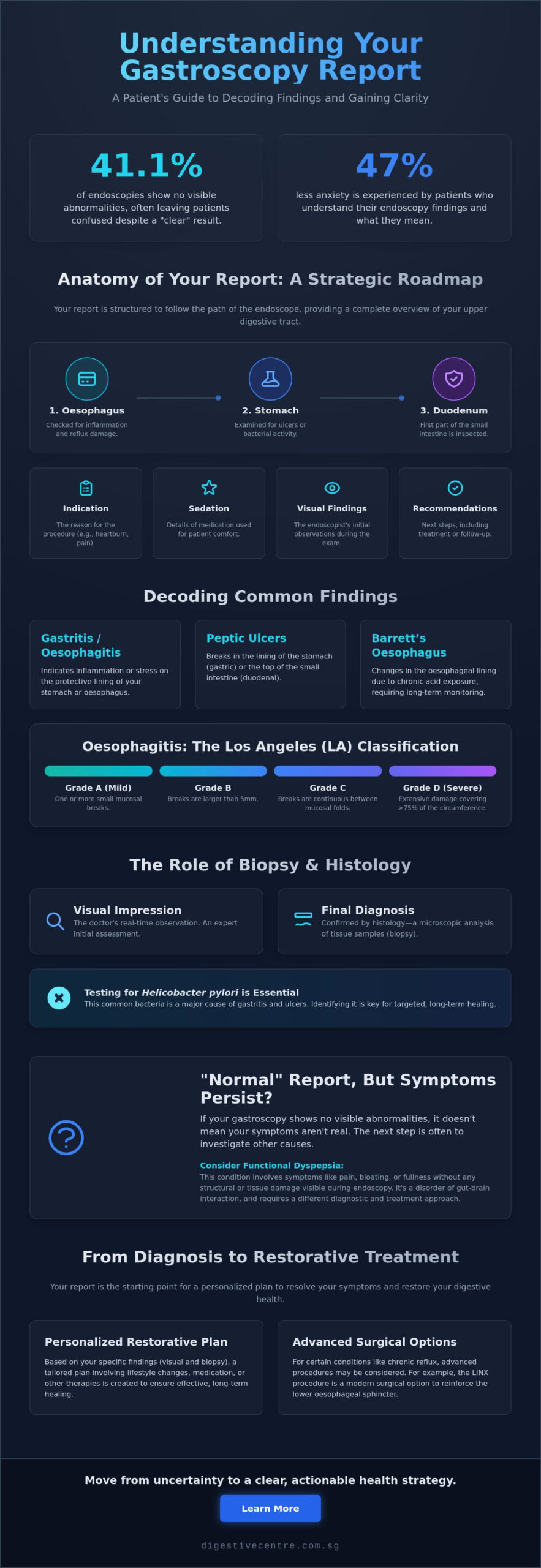
Did you know that approximately 41.1% of diagnostic endoscopies show no visible abnormalities, yet many patients still leave the clinic feeling more confused than when they arrived? It’s completely natural to feel a surge of anxiety when you’re handed a document filled with complex clinical terms. You want answers about your digestive health, but the process of interpreting your gastroscopy report can feel like learning a foreign language. This confusion often leads to unnecessary stress, especially when you’re waiting for biopsy results or wondering why your symptoms persist despite a seemingly clear report.
Research indicates that patients who understand their endoscopy findings experience 47% less procedure-related anxiety. This guide simplifies the clinical jargon, giving you the clarity you need to understand your findings and what they mean for your long-term recovery. We’ll break down the structure of your medical report and map out a logical path toward resolving your symptoms. By the end of this article, you’ll have a professional perspective on your results, ensuring you move forward with confidence and the peace of mind that comes from true understanding.
Key Takeaways
- Navigate the professional structure of your medical documentation to understand how clinical indications shape your final diagnosis.
- Gain confidence in interpreting your gastroscopy report by decoding common findings like gastritis grades and the structural impact of a hiatus hernia.
- Understand the vital role of histology and why testing for Helicobacter pylori is essential for targeted, long-term healing.
- Learn why a “normal” visual finding may still require investigation into functional dyspepsia to resolve ongoing symptoms effectively.
- Discover how to transition from a diagnostic report to a personalized restorative plan, including advanced surgical options like the LINX procedure.
Understanding the Anatomy of Your Gastroscopy Report
Your medical report might look like a wall of clinical text, but it follows a highly logical sequence designed to provide a comprehensive overview of your upper digestive tract. A gastroscopy report is a strategic roadmap. It documents every millimeter of your upper gastrointestinal tract to ensure no detail is overlooked. When you begin interpreting your gastroscopy report, it’s helpful to recognize that the document is divided into distinct, functional zones. These zones typically include the indication for the procedure, the sedation details, the visual findings across three anatomical regions, and the final recommendations for your care.
The report is structured to follow the physical path of the endoscope. It begins with the oesophagus, where the specialist looks for signs of inflammation or reflux damage. Next, it moves to the stomach to check for ulcers or bacterial activity. Finally, it reaches the duodenum, which is the first part of the small intestine. To better understand the scope of this examination, one might ask, What is a Gastroscopy (EGD)? It is a diagnostic tool that relies on high-definition imaging to detect subtle mucosal changes. These advanced optics allow for earlier intervention and more accurate restorative plans, ensuring your long-term health is prioritized.
The Clinical Context: Indication and Sedation
The “Indication” section is a critical part of the document. It lists the specific reasons for your referral, such as persistent heartburn, unexplained weight loss, or chronic abdominal pain. This context acts as a lens through which the endoscopist views your anatomy. If your indication is chronic reflux, the specialist will pay closer attention to the gastro-oesophageal junction. Regarding your comfort, most procedures in Singapore utilize “conscious sedation.” This typically involves a combination of sedative and analgesic medications. It ensures you remain relaxed and pain-free while allowing the specialist to conduct a thorough and efficient examination.
Visual Findings vs. Final Diagnosis
It’s vital to distinguish between what the doctor sees and what the lab eventually confirms. Immediately after the procedure, the specialist records their visual “impression.” This is an expert observation based on real-time footage. However, a visual finding of “gastritis” is often an initial assessment. A final, definitive diagnosis usually requires histology, where tissue samples are analyzed under a microscope. Your report will include high-resolution photographic evidence documenting your digestive health. These images provide a baseline for tracking your recovery and ensure that your path toward wellness is based on objective, visible data.
Decoding Common Findings: Gastritis, Ulcers, and Reflux
When you move beyond the anatomical labels, your report begins to describe the health and vitality of your digestive lining. This is where interpreting your gastroscopy report becomes highly personalized. Findings like gastritis or oesophagitis indicate that the protective layers of your stomach or food pipe are under stress. While these terms sound clinical, they are essentially a snapshot of inflammation that allows us to build an efficient recovery plan. Understanding the specific nature of these findings, such as whether they are erosive or non-erosive, helps determine how quickly you can expect to return to peak physical comfort.
Peptic ulcers are another common discovery, often categorized by their location. Gastric ulcers occur in the stomach lining, while duodenal ulcers are found at the beginning of the small intestine. The severity of these ulcers dictates the intensity of the restorative approach. In some cases, the specialist may observe “tongues” of red, velvety tissue extending upward from the stomach. This is often an indicator of Barrett’s oesophagus, a condition where the lining changes due to chronic acid exposure. Identifying this early is a proactive step that ensures long-term monitoring and safety. Receiving your gastroscopy results is the first step in moving from uncertainty to a clear, actionable health strategy.
Inflammation Grades and Severity
Oesophagitis is typically measured using the Los Angeles (LA) Classification, which ranks inflammation from Grade A (mild) to Grade D (severe). Grade A involves small mucosal breaks, while Grade D indicates extensive damage involving a large portion of the circumference. Understanding these grades is essential for tracking your treatment timeline. Severe inflammation often correlates with specific signs you need an urgent gastroscopy, ensuring that interventions are timed for maximum efficacy and restorative benefit.
Structural Findings: Hernias and Strictures
Structural changes often explain why symptoms persist despite medication. A hiatus hernia, where the stomach pushes through the diaphragm, is a primary driver of chronic acid reflux. Sliding hernias are common and may move in and out of the chest, while para-oesophageal hernias are more fixed and often require a precise surgical approach. Your report may also mention “Z-line” irregularities, which mark the junction where the oesophagus meets the stomach. If this line is blurred or irregular, it suggests that chronic reflux has begun to alter the local tissue environment. If structural issues are identified, exploring advanced reflux treatments can help restore your digestive efficiency and quality of life.
The Biopsy Report: Understanding Histology and Cell Changes
While a visual inspection provides immediate feedback, the biopsy report offers the definitive microscopic detail required for a precise diagnosis. It’s the most scientific part of interpreting your gastroscopy report. A biopsy, or histology, involves taking tiny tissue samples to examine cell structure and health under a microscope. This isn’t just about looking for serious illnesses. It’s about identifying the root causes of inflammation and preventing future complications before they start. By analyzing these samples, your specialist can determine exactly why your digestive system isn’t performing at its peak.
When you are interpreting your gastroscopy report, remember that these histology results provide the scientific foundation for your next steps. What your biopsy results mean in this context is often the difference between simply managing symptoms and achieving a permanent cure. These findings allow for a tailor-made treatment plan that addresses your specific cellular health, ensuring a faster return to your daily routine.
Screening for H. Pylori and Infections
One of the most common reasons for a biopsy is to check for Helicobacter pylori (H. pylori). This bacterium is a primary cause of chronic gastritis and peptic ulcers. During the procedure, your specialist may use a CLO test, also known as a Rapid Urease Test. This provides a nearly immediate result by detecting an enzyme produced by the bacteria. If the test is positive, a targeted course of antibiotics and acid-suppressing medication is prescribed. Successfully treating this infection is vital for your long-term health. It prevents the recurrence of ulcers and significantly reduces the risk of more serious stomach conditions. After the initial treatment, follow-up tests are often scheduled to confirm the bacteria have been fully eradicated.
Metaplasia and Dysplasia Explained
Hearing terms like “metaplasia” or “dysplasia” often causes anxiety, but these findings are manageable when caught early. Intestinal Metaplasia is a common adaptive change. It means the stomach lining has replaced some of its cells with cells that look more like those in the intestine. This is usually a response to chronic irritation, such as long-term acid reflux. Dysplasia represents a more significant shift in cell growth. Low-grade dysplasia requires regular surveillance to ensure the cells don’t progress. High-grade dysplasia is more serious and often necessitates proactive intervention. Biopsies also play a critical role in ruling out malignancy in chronic gastric ulcers, providing a safety net for your health. Additionally, findings like atrophic gastritis indicate a thinning of the stomach lining, which can affect how your body absorbs essential nutrients like Vitamin B12 and iron.
Beyond the Paper: When Results are Normal but Symptoms Persist
Receiving a “macroscopically normal” report can be a confusing experience when you’re still dealing with daily discomfort. Approximately 41.1% of diagnostic endoscopies show no visible abnormalities, but this doesn’t mean your symptoms aren’t real. When interpreting your gastroscopy report, a normal result simply confirms that the structure of your organs is intact and free of visible lesions, ulcers, or tumors. It marks the transition from looking for structural damage to investigating functional and metabolic efficiency. This is a critical distinction that moves your care toward more sophisticated diagnostic territory.
In many cases, persistent symptoms like bloating or upper abdominal pain are caused by functional dyspepsia. This is a condition where the stomach looks perfectly healthy under high-definition imaging but doesn’t function correctly in terms of movement or sensitivity. Similarly, Silent Reflux (LPR) can often be missed during a standard resting gastroscopy because the damage may be microscopic or occur only during specific activities. If your report is clear but your quality of life is still affected, consulting unexplained abdominal pain specialists is the logical next step to solve the paradox of chronic discomfort.
Functional vs. Structural Issues
A clear gastroscopy focuses on anatomy, but it doesn’t always capture gastric motility. You might have a “normal” stomach that simply empties too slowly, leading to significant bloating and early fullness. There’s also a deep connection between gut health and metabolic conditions like Polycystic Metabolic Overgrowth Syndrome (PMOS). If interpreting your gastroscopy report leaves you with more questions than answers, your specialist may recommend follow-up tests like pH monitoring or manometry. These tools measure the actual performance and acid exposure levels of your digestive tract over a 24-hour period, providing data that a visual scope cannot see.
Metabolic and Lifestyle Correlations
Your digestive efficiency is often a reflection of your overall metabolic health. High levels of visceral fat and systemic inflammation can put physical pressure on the stomach, mimicking the symptoms of reflux even when the lining looks healthy. Insulin resistance is another factor that can slow down digestive transit time, leading to a heavy, uncomfortable feeling after meals. Nutritional counselling is often the missing piece of the puzzle for patients with “normal” reports. By addressing the underlying metabolic drivers of your discomfort, you can achieve restorative benefits that medication alone cannot provide. If you’re ready to look deeper into these connections, a comprehensive metabolic health assessment can help align your digestive function with your long-term wellness goals.
Navigating Next Steps: From Diagnosis to Restorative Treatment
Once you have finished interpreting your gastroscopy report, the focus shifts from data collection to active, personalized healing. A medical report isn’t a final destination. It’s the essential foundation for a restorative plan designed to return you to peak physical comfort. Whether your results showed specific inflammation, structural changes, or even “normal” findings paired with persistent symptoms, the next phase of your journey is about precision. We move beyond simply documenting illness and begin the process of restoring your digestive and metabolic efficiency through a multi-disciplinary approach.
This transition is where modern specialized care excels. By using the high-definition data from your procedure, we can bypass the trial-and-error often associated with general digestive care. Your path forward might involve advanced endoscopic interventions, surgical barrier restoration, or metabolic optimization. The objective is to create a seamless, high-performance journey that prioritizes your long-term well-being and minimizes the time you spend managing symptoms. Interpreting your gastroscopy report with a specialist ensures that every finding is mapped to a tangible, restorative benefit.
Advanced Interventions for Chronic GERD
Many patients find themselves reliant on daily acid-suppressing medications for years without achieving true resolution. When your gastroscopy confirms structural reflux or a weak lower oesophageal sphincter, moving beyond medication is often the most effective path. The LINX system is a sophisticated, minimally invasive solution that uses a small ring of magnetic beads to restore the natural barrier of the oesophagus. This intervention is particularly vital if your report identified early signs of Barrett’s Oesophagus. By restoring the physical barrier, we can prevent the progression of cell changes and provide a permanent solution to chronic reflux damage.
Integrated Metabolic and Digestive Care
Your digestive health is deeply connected to your metabolic profile. If your findings suggest that weight or systemic inflammation is driving your GI distress, integrated medical weight management can be a transformative step. Generic medications such as semaglutide or tirzepatide are often utilized to support this process, helping to reduce visceral fat and improve overall GI transit times. For individuals requiring more significant metabolic intervention, Endoscopic Sleeve Gastroplasty (ESG) offers a non-surgical, suture-based method to reduce stomach volume and improve metabolic efficiency.
True recovery also requires a focus on the underlying drivers of your health. Metabolic testing and professional nutritional counselling provide the data and guidance needed to ensure your results are sustainable. This holistic destination ensures that every aspect of your health, from cellular changes to lifestyle habits, is addressed. Scheduling your follow-up consultation is the final, frictionless step in your journey, ensuring your path to recovery is handled with professional speed and expert care.
Restoring Your Digestive Efficiency and Long-Term Health
Your medical documentation is more than a clinical record; it’s a strategic roadmap for restoring your digestive and metabolic efficiency. By moving beyond the initial confusion of medical jargon, you gain the clarity needed to make informed decisions about your recovery. Interpreting your gastroscopy report with a professional perspective allows you to address the root causes of your symptoms, whether they are structural, functional, or metabolic in nature.
At the Digestive Centre, we specialize in bridging the gap between advanced diagnostics and restorative results. Led by Senior Consultant Surgeon Dr. Shanker Pasupathy, our team integrates comprehensive metabolic testing with cutting-edge interventions such as the LINX procedure and Endoscopic Sleeve Gastroplasty (ESG). We prioritize accelerated timelines and reduced discomfort to help you return to your peak performance as quickly as possible. Schedule a consultation with Dr. Shanker Pasupathy to discuss your results and take the final step toward lasting digestive wellness. Your path to a healthier, more comfortable life begins with a clear understanding of your body’s unique needs.
Frequently Asked Questions
How long does it take to get the final biopsy results after a gastroscopy?
Final biopsy results typically take between three to seven working days to process in Singapore. This timeline allows the pathologist to carefully examine tissue samples for cellular changes or infections like H. pylori. Once the lab completes its analysis, your specialist will review the findings to finalize your restorative plan. This ensures your treatment is based on precise microscopic data rather than just visual impressions.
What does “erythematous mucosa” mean in my gastroscopy report?
“Erythematous mucosa” is a clinical term describing redness in the lining of your stomach or oesophagus. This redness is a visual indicator of inflammation, often caused by acid irritation, bile reflux, or infections. While it sounds complex, it’s a common finding when interpreting your gastroscopy report. It signals that the protective layer of your digestive tract is under stress and requires targeted care to restore its health.
If my gastroscopy is normal, why do I still have chronic acid reflux?
A normal gastroscopy confirms there’s no visible structural damage, but it doesn’t rule out functional issues. You may have Non-Erosive Reflux Disease (NERD), where acid causes symptoms without leaving visible scars. In these cases, your specialist might recommend pH monitoring or manometry. These advanced tests measure the actual performance of your oesophageal valve, helping to solve the paradox of persistent symptoms despite a clear visual report.
Does a hiatus hernia found during gastroscopy always require surgery?
A hiatus hernia doesn’t always require surgical intervention. Small, sliding hernias are often managed through lifestyle adjustments and medication to control acid production. However, if the hernia is large or causing severe, persistent reflux that impacts your quality of life, surgical solutions like the LINX procedure might be recommended. The decision depends on the structural severity documented in your report and how well your symptoms respond to conservative restorative treatments.
What is the difference between a gastroscopy report and a histology report?
The gastroscopy report documents what the specialist sees during the procedure, while the histology report describes what the pathologist sees under a microscope. Think of the gastroscopy report as a visual map and the histology report as a detailed cellular analysis. Both are essential for interpreting your gastroscopy report accurately. Together, they provide the clinical authority needed to confirm diagnoses like bacterial infections or early-stage cell changes.
Can a gastroscopy miss certain conditions like silent reflux?
Yes, a standard gastroscopy can sometimes miss “silent reflux” (LPR) because the procedure is a resting snapshot of your anatomy. LPR often affects the throat and vocal cords, areas that may not show significant damage during a routine upper GI scope. If symptoms like a chronic cough or throat clearing persist despite a normal report, functional testing or metabolic assessments are often the next logical steps to identify the root cause.
What should I do if my report mentions “Barrett’s Oesophagus”?
If your report mentions Barrett’s Oesophagus, the primary goal is proactive monitoring and reflux control. This finding means the lining of your food pipe has changed due to chronic acid exposure. Your specialist will typically recommend a regular surveillance schedule to check for cell progression. Managing the underlying reflux through advanced interventions or lifestyle changes is vital to protect your long-term health and prevent further cellular shifts from occurring.
Is it normal to have a sore throat or bloating after the procedure?
It’s completely normal to experience a mild sore throat or temporary bloating immediately after the procedure. The sore throat is usually caused by the passage of the endoscope, while bloating results from the air used to expand the stomach for better visibility. These minor discomforts are temporary and typically resolve within 24 hours. Most patients find that their physical comfort returns quickly, allowing them to resume their normal daily activities almost immediately.